2.29. DSA Mode
Using the DSA mode you can process multiphase and multiframe series with a varying concentration of a contrast agent, subtracting images in such a way that the tissues containing contrast are best seen. Applicable to series with CT, MRI and X-ray modalities.
The following outlines how to view multiframe series. Viewing multiphase series of studies is done in the same way.
 | Hight-density tissue (e.g. bone) images may be subtracted from the images of contrast-enhanced vessels, which will lead to data loss. |
2.29.1 Single-image subtraction view
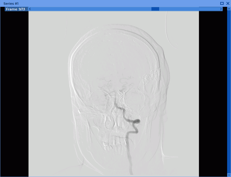
For studies containing multiframe series with a varying concentration of a contrast agent, one of the frames is subtracted from all frames in the series. To apply the DSA mode:
-
Open the series of study in the View Flat Images window.
-
Click on the Digital Subtraction Angiography (DSA)
 button. The first frame
of the series has been subtracted from all the frames of this series.
button. The first frame
of the series has been subtracted from all the frames of this series.
-
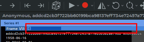
Scroll through the frames by moving the slider or rotating the mouse wheel, hovering the cursor over the scroll bar on the Frame Scroll Bar (Fig. 2.89).
-
If you want to subtract another frame from all the frames, select this image using the scroll bar in the Frame Scroll Bar, click on the arrow on the
button and select the
command Set the current frame as a key frame.
-
If the series frames are offset relative to each other, then correct it by selecting the command Auto adjust key image offset from the Digital Subtraction Angiography (DSA) tool menu.
-
To manually correct the offset, select the command Manual Adjustment from the Digital Subtraction Angiography (DSA) tool menu and change the location of the frames using the buttons that appear in the image view window (Fig. 2.90) or using the


 keys.
keys.
-
If necessary, change the window width and the window level.
2.29.2 Series subtraction view
For studies containing at least two related series of frames and images with different concentrations of a contrast agent, the frames and images of one series are subtracted from the corresponding frames and images of the other series. To apply the DSA mode:
-
Open the series of study in the View Flat Images window.
-
Choose a series whose images you want to subtract from the images of the other series.
-
Click on the
button.
-
Click on the arrow on the Digital Subtraction Angiography (DSA)
button.
If the series contains the necessary data, then the command Set the frame series as
key frames is available. Select this command.
-
Select another series of frames by rotating the mouse wheel or moving the slider on the right side of the image view window.
-
Scroll through the images by moving the slider or rotating the mouse wheel, hovering the cursor over the scroll bar on the Frame Scroll Bar (Fig. 2.89).
-
If necessary, change the window width and the window level.
This tool is also available from the Image main menu item.
2.29.3 Viewing Several Series of the Same Study Fused in One Multiphase Series with Subtraction
A fused series may be viewed in the digital subtraction angiography (DSA) mode and used for assessment of contrast distribution in tissues at regular time intervals.
-
Open a fused series in the flat image viewer mode (see Section 2.12).
-
Scroll the phases and images of the series by moving the sliders on the phases and images scroll bars.
-
Activate the DSA mode by clicking the Digital Subtraction Angiography (DSA)
button.
-
Chose a phase whose images are to be subtracted from the images belonging to other phases. Click the arrow on the
button and select the Set the current frame as
a key frame option on the tool menu.
-
Change the window width and level if required.
This tool is also available from the Image main menu item.