9.3. MIP mode
____________________________________________________________________________________________
Functionality is available in a separate module which is activated in the Pro edition for an extra fee
____________________________________________________________________________________________
The DICOM Viewer allows you to change the section thickness for flat images, i.e. the image is actually shown in MIP (Maximum Intensity Projection) mode. To change the section thickness, you need to sort the images by phases and frames (the default setting for the sorting procedure; for details see Section 2.1). The changes are possible within 0-50 mm range. You cannot make the section thinner than the source image.
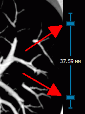
To change the thickness, use the tool shown in Figure 9.4, which you will find on the right-hand side of the window in Mono-CT, Mono-PET and Fusion PET+CT modes (see Section 9.2). In the centre you can see the current section thickness. The sliders adjusting the thickness are marked with arrows. To change the thickness, you need to move either of the sliders by pressing the left, right or middle mouse button. To make the section thicker, move the slider outwards, to make it thinner — inward.
The thickness set with the help of the tool is used when exporting and adding to the print list.
When you are working with such tools as Point value, ROI rectangle, ROI ellipse and ROI polygon, the data shown are for MIP mode images. When the thickness is changed, the value is automatically scaled. All the other measurements are performed for the source data.